
Dr. Jiung Cho and dr. Miri Choi of KBSI imaged Magnetosprilum Magnetotacticum (MSR-1) bacteria from KIST Europe in their advanced TEM system. The results are exiting. By negative staining, they succeeded in imaging the flagella really clearly. The high resolution images of the magneto-some crystals very beautifully show the Fe3O4 FCC crystal structure.
For transmission electron microscopy (TEM) analysis, the medium with MSR-1 MTB was applied on carbon coated copper grids (CF200-Cu) and allow to absorb for 30s. Excess sample was blotted off by touching the edge of the grid with clean piece of filter paper and stained with 2% uranyl acetate solution for 30 s.The morphology of MTB was examined by a JEM-2100F TEM (JEOL, Japan) with bright field image at an accelerating voltage of 200 kV.
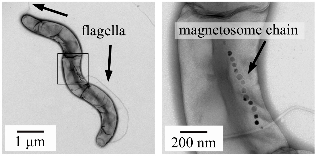
Figure 1: Bright field transmission electron microscope images of the MRS-1 magneto-tactic bacteria. In this negatively stained image, the flagella can be clearly observed (left) as well as the magnetosome chain (right).
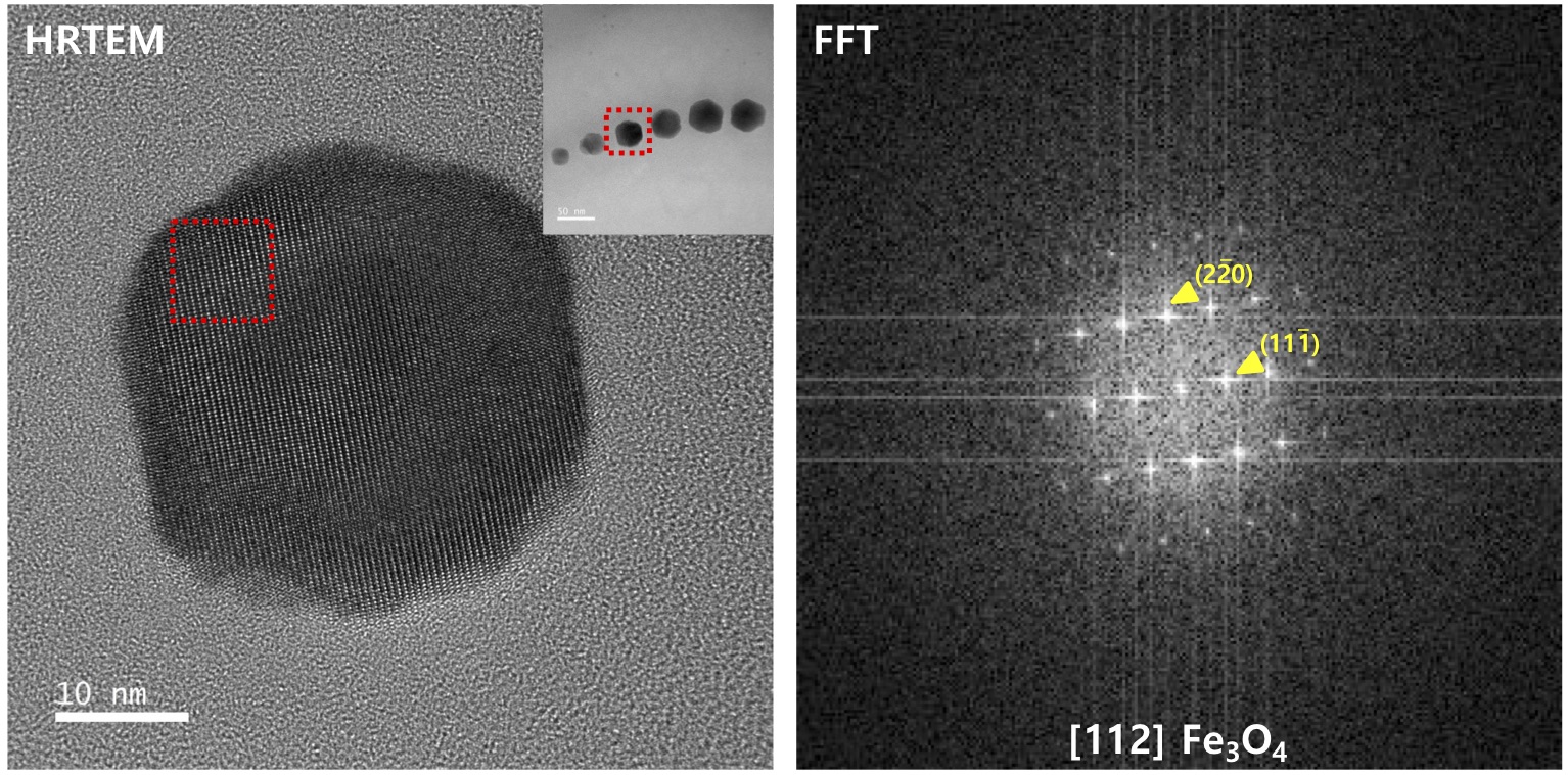
Figure 2: Bright field transmission electron microscope images (left) and FFT extracted diffraction pattern (Right). The pattern clearly indicates an FCC Fe3O4 structure