

We are proud to announce that Yonsei University has joined our consortium. The IBS research team of professor Jinwoo Cheon has been working for a long time with dr. Damien Faivre of MPI and CEA. Their expertise will strengthen the consortium in the application of magnetic nanoparticles and magneto-tactic bacteria in biomedicine.

Dr. Damien Faivre of CEA, Cadarache, visited KIST Europe to prepare for the upcoming Korea-EU workshop. We took advantage of the visit of this outstanding expert in the field of magneto-tactic bacteria, and made him present his work for the Saarland research community.
In november Prof. Abelmann of KIST Europe brought MSR-1 bacteria to DGIST. Dr. Jin-Young Kim of the team of prof. Hongsoo Choi and dr. Thomas John of Saarland University succeeded in magnetic control of the bacteria in the DGIST advanced microscope setup. Below is a snapshot of a video recording of an experiment during which a rotating magnetic field is switched on and off. When on, the fraction of MSR-1 that are magnetic follow the magnetic field.
Snapshot of a video in which MSR-1 bacteria are controlled. When the field is ON (top line), the fraction of MSR-1 that are magnetic follow the rotating magnetic field.
Dr. Jiung Cho and dr. Miri Choi of KBSI imaged Magnetosprilum Magnetotacticum (MSR-1) bacteria from KIST Europe in their advanced TEM system. The results are exiting. By negative staining, they succeeded in imaging the flagella really clearly. The high resolution images of the magneto-some crystals very beautifully show the Fe3O4 FCC crystal structure.
For transmission electron microscopy (TEM) analysis, the medium with MSR-1 MTB was applied on carbon coated copper grids (CF200-Cu) and allow to absorb for 30s. Excess sample was blotted off by touching the edge of the grid with clean piece of filter paper and stained with 2% uranyl acetate solution for 30 s.The morphology of MTB was examined by a JEM-2100F TEM (JEOL, Japan) with bright field image at an accelerating voltage of 200 kV.
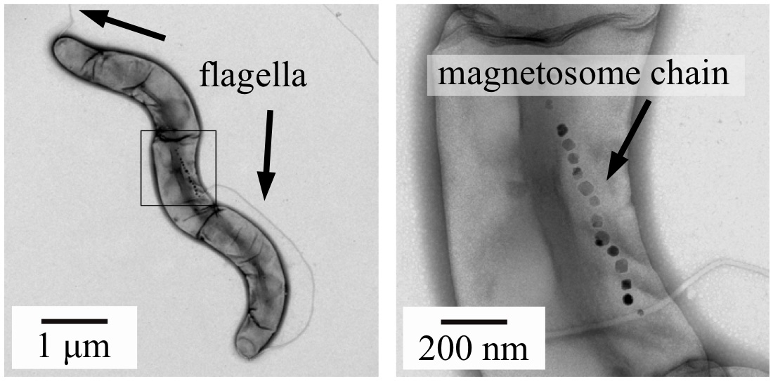
Figure 1: Bright field transmission electron microscope images of the MRS-1 magneto-tactic bacteria. In this negatively stained image, the flagella can be clearly observed (left) as well as the magnetosome chain (right).
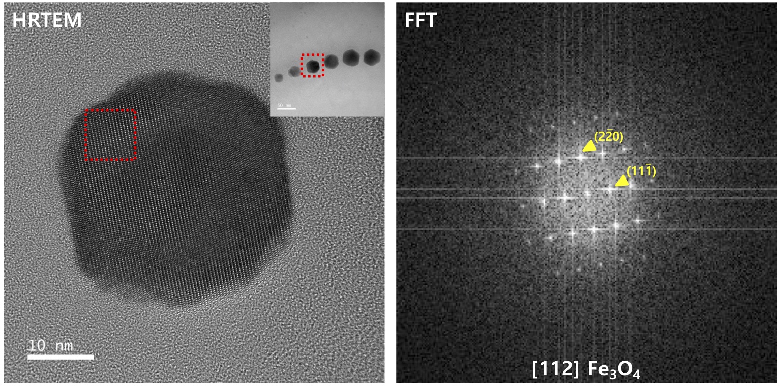
Figure 2: Bright field transmission electron microscope images (left) and FFT extracted diffraction pattern (Right). The pattern clearly indicates an FCC Fe3O4 structure
Page 2 of 4
- You are here:
-
Home

- News